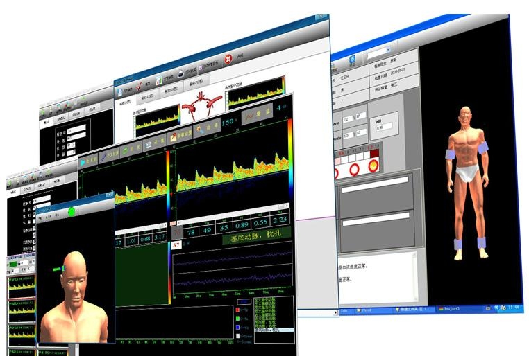

The correlation between invasive and non-invasive ICP measurements was good (R = 0.74), and the 95 % limits of agreements were -1.4 ± 8.8 mmHg. Thus, twelve patients and a total of 61 paired data points were studied. An infusion test could not be performed in one patient. Reliable Doppler signals were obtained in 13 (72 %) patients. Bench tests of the infusion apparatus showed a random error (95 % CI) of less than ☐.9 mmHg and a systematic error of less than ☐.5 mmHg. Ringer's solution was used to create elevation to pre-defined ICP levels. As a reference, an automatic CSF infusion apparatus was connected to the lumbar space. Koskinen et al performed multiple measurements over a wide ICP span in eighteen elderly patients with communicating hydrocephalus. The aim of a study was to investigate how well non-invasively-measured ICP and invasively-measured cerebrospinal fluid (CSF) pressure correlate. This method is further developed by Company Vittamed (Kaunas, Lithuania) together with consortium partners in EU FP7 project Brainsafe ) As a result of that all individual influential factors (ABP, cerebrovascular autoregulation impairment, individual pathophysiological state of patient, individual diameter and anatomy of OA, hydrodynamic resistance of eye ball vessels, etc.) do not influence the balance aICP=aPe and, as a consequence, such natural “scales” do not need calibration. The mean value of OA blood flow, it’s systolic and diastolic values, pulsatility indexes are almost the same in both OA segments in the point of balance aICP=aPe. This measurement method eliminates the main limiting problem- the individual patient calibration problem by direct comparison of aICP and externaly applied pressure – same fundamental principle used to measure blood pressure with a sphygmomanometer. At this point, the applied external pressure equals the intracranial pressure. The aICP meter based on this method gradually increases the pressure over the eye so that the blood flow parameters in two sections of artery are equal. In place of the stethoscope, a Doppler ultrasound beam measures the blood flow in intracranial and extracranial segments of the Ophthalmic Artery. Blood flow in the intracranial segment is affected by intracranial pressure, while flow in the extracranial segment is influenced by the externally applied pressure to the orbital tissues.Īs with a sphygmomanometer, a pressure cuff is used - in this case to compress the tissues surrounding the eye and change the characteristics of blood flowing from inside the skull cavity into the eye socket. Ophthalmic artery (OA) - a unique vessel with intracranial and extracranial segments is used as a natural pair of scales for absolute ICP measurement. along the entire course of the MCA and PCA and at 2 depths from the ACA and distal. The TDTD method uses Doppler ultrasound to translate principle of blood pressure measurement with a sphygmomanometer to the measurement of ICP. Transcranial Doppler ultrasound (TCD) is a noninvasive technique that. Innovative method using Two-Depth Transcranial Doppler (TDTD) of monitoring intracranial pressure (ICP) relies on the same fundamental principle used to measure blood pressure with a sphygmomanometer. To avoid lumbar puncture or intracranial ICP probes, non-invasive ICP techniques are becoming popular. From these hemodynamic changes, transcranial Doppler sonographic diagnostic criteria for MCA occlusive and stenotic lesions were established.Measurement of intracranial pressure (ICP) is necessary in many neurological and neurosurgical diseases. If there was a high-grade stenosis or occlusion of the internal carotid artery, a collateral circulation over the anterior part of the circle of Willis was seen in addition to the changes caused by the MCA disease. In MCA stenosis a steep rise of MCA FV appeared inside the stenotic segment. Central MCA lesions showed less marked changes than did peripheral lesions. At the same time the anterior cerebral artery FV increased because of collateral flow over leptomeningeal anastomoses. With MCA lesions, the MCA flow velocity (FV) was reduced. According to computed tomographic, angiographic, and/or autopsy findings, the patients were classified as having MCA occlusive lesions in the central (sphenoidal) part or in peripheral branches or MCA stenosis. The transcranial Doppler sonographic findings of 61 patients with middle cerebral artery (MCA) disease were compared with those of 535 controls.


 0 kommentar(er)
